


 Limited Edition Golden Llama is here! Check out how you can get one.
  Limited Edition Golden Llama is here! Check out how you can get one.
 Offering SPR-BLI Services - Proteins provided for free!
  Offering SPR-BLI Services - Proteins provided for free!
 Thank you for choosing ACROBiosystems. Would you rate our product and service?
  Thank you for choosing ACROBiosystems. Would you rate our product and service?
 Here come GMP Grade Cytokines!Free Sample is available!
  Here come GMP Grade Cytokines!Free Sample is available!
> Neural Antibodies | The Key to Precise Detection of Nerve Cell Markers

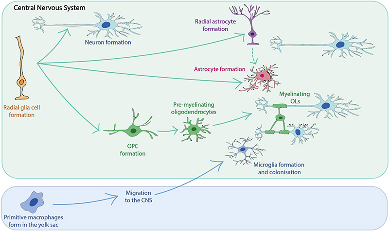
| Molecule | Cat. No. | Product Description | Application |
|---|---|---|---|
| GFAP | GFP-S453 | Polyclonal GFAP Antibody, Rabbit IgG | Specific binding with GFAP to identify astrocytes |
| NG2/Cspg4 | NG4-S455 | Polyclonal NG2/Cspg4 Antibody, Rabbit IgG | Specific binding with NG2 to identify oligodendrocyte progenitors |
| Olig2 | OL2-S456 | Polyclonal Olig2 Antibody, Rabbit IgG | Specific binding with Olig2 to identify oligodendrocytes |
| Tbr1 | TB1-S457 | Polyclonal Tbr1 Antibody, Rabbit IgG | Specific binding with Tbr1 to identify immature neurons |
| NeuN/Rbfox3 | NE3-S454 | Monoclonal NeuN/Rbfox3 Antibody, Mouse IgG1 | Specific binding with NeuN to identify mature neurons |
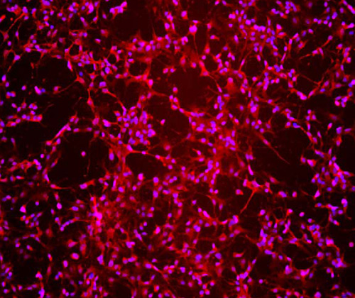
Immunofluorescent staining (10X) of cerebral organoid-derived cells (Cat. No. CIPO-BWL001K) labeling Tbr1 (Red) with purified Polyclonal Tbr1 Antibody, Rabbit IgG (Cat. No. TB1-S457) at 1:200 dilution. DAPI (blue) was used as nuclear counterstain.
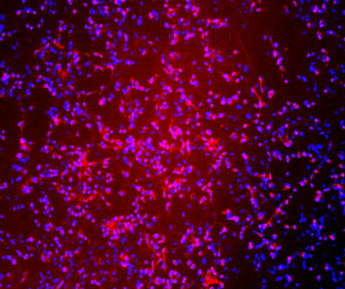
Immunofluorescent staining (10X) of cerebral organoid-derived cells (Cat. No. CIPO-BWL001K) labeling NeuN (Red) with purified Monoclonal NeuN/Rbfox3 Antibody, Mouse IgG1 (Cat. No. NE3-S454) at 1:500 dilution. DAPI (blue) was used as nuclear counterstain.

Immunofluorescent staining (10X) of cerebral organoid-derived cells (Cat. No. CIPO-BWL001K) labeling GFAP (Red) with purified Polyclonal GFAP Antibody, Rabbit IgG (Cat. No. GFP-S453) at 1:200 dilution. DAPI (blue) was used as nuclear counterstain.
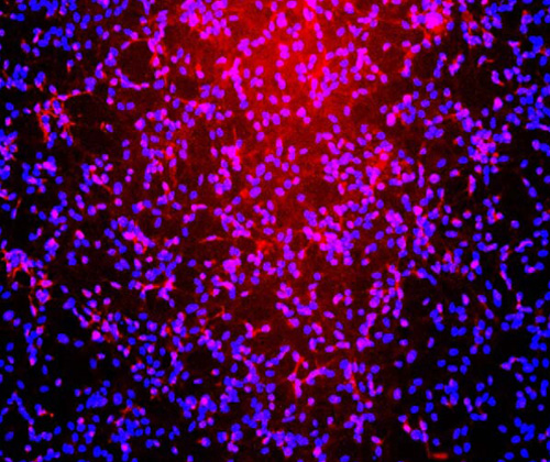
Immunofluorescent staining (10X) of cerebral organoid-derived cells (Cat. No. CIPO-BWL001K) labeling NG2 (Red) with purified Polyclonal NG2/Cspg4 Antibody, Rabbit IgG (Cat. No. NG4-S455) at 1:200 dilution. DAPI (blue) was used as nuclear counterstain.
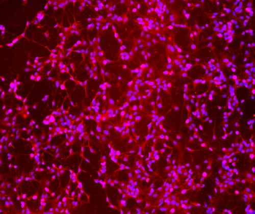
Immunofluorescent staining (10X) of cerebral organoid-derived cells (Cat. No. CIPO-BWL001K) labeling Olig2 (Red) with purified Polyclonal Olig2 Antibody, Rabbit IgG (Cat. No. OL2-S456) at 1:200 dilution. DAPI (blue) was used as nuclear counterstain.
1. Neely S A, Lyons D A. Insights into central nervous system glial cell formation and function from zebrafish[J]. Frontiers in Cell and Developmental Biology, 2021, 9: 754606. https://doi.org/10.3389/fcell.2021.754606
2. Yokoo H, Nobusawa S, Takebayashi H, et al. Anti-human Olig2 antibody as a useful immunohistochemical marker of normal oligodendrocytes and gliomas[J]. The American journal of pathology, 2004, 164(5): 1717-1725. https://doi.org/10.1016/S0002-9440(10)63730-3
3. Zhao W, Dumanis S B, Tamboli I Y, et al. Human APOE genotype affects intraneuronal Aβ1–42 accumulation in a lentiviral gene transfer model[J]. Human molecular genetics, 2014, 23(5): 1365-1375. https://doi.org/10.1093/hmg/ddt525
4. Yu L, Chen C, Wang L F, et al. Neuroprotective effect of kaempferol glycosides against brain injury and neuroinflammation by inhibiting the activation of NF-κB and STAT3 in transient focal stroke[J]. PloS one, 2013, 8(2): e55839. https://doi.org/10.1371/journal.pone.0055839
5. Rigo Y R, Benvenutti R, Portela L V, et al. Neurogenic potential of NG2 in neurotrauma: a systematic review[J]. Neural Regeneration Research, 2024, 19(12): 2673-2683. https://doi.org/10.4103/NRR.NRR-D-23-01031
6. Englund C, Fink A, Lau C, et al. Pax6, Tbr2, and Tbr1 are expressed sequentially by radial glia, intermediate progenitor cells, and postmitotic neurons in develo** neocortex[J]. Journal of Neuroscience, 2005, 25(1): 247-251. https://doi.org/10.1523/JNEUROSCI.2899-04.2005
7. Jurga A M, Paleczna M, Kadluczka J, et al. Beyond the GFAP-astrocyte protein markers in the brain[J]. Biomolecules, 2021, 11(9): 1361. https://doi.org/10.3390/biom11091361
8. Alekseeva O S, Gusel’nikova V V, Beznin G V, et al. Prospects for the application of neun nuclear protein as a marker of the functional state of nerve cells in vertebrates[J]. Journal of Evolutionary Biochemistry and Physiology, 2015, 51: 357-369. https://doi.org/10.1134/S0022093015050014
This web search service is supported by Google Inc.


